By: solteszlab

October 13, 2020

Patch-Clamp Recordings

The brain is composed of a wide array of interconnected neurons, including both excitatory principal cells and a diverse assortment of regulatory interneurons. As neurons communicate through electrochemical mechanisms, characterizing the electrical signatures of both control and experimentally perturbed neurons provides great insight into the properties of the fundamental components that compose brain circuits.

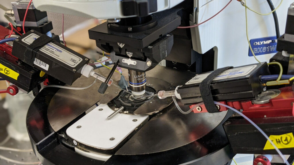
With whole cell electrophysiology, we use finely pointed glass electrodes to precisely record the electrical signal from within individual neurons. Thus, we can examine both the functional electrical properties intrinsic to that particular cell and monitor the synaptic inputs that neuron receives. While recordings are often made from the neuronal cell body, the Soltesz lab has experience recording specifically from smaller structures, including hippocampal dendrites or even axonal terminals.

We also utilize several variants on the whole cell electrophysiology approach to interrogate other features of neuronal networks. We can apply compounds to manipulate neuronal activity either through bath application onto the entire brain slice, or through more targeted application via a microinjection system. Through paired patch-clamp recordings, we explore how two simultaneously recorded neurons (whether of the same or different cell types) are interconnected and how they influence the electrical properties of each other. We also employ optogenetic techniques to alter the activity of genetically targeted neuronal populations in response to light stimulation, both to examine their downstream connectivity and how they influence the electrical properties of other neurons. Furthermore, we have the capacity to combine whole cell electrophysiology with simultaneous high-speed imaging of optical probes (e.g. calcium and voltage indicators), enabling us to incorporate the high single cell precision of electrophysiology with broader population level readouts of activity within a brain slice.

Neurons recorded with whole cell electrophysiology can be filled with intracellular tracers, allowing enabling later morphological reconstruction of the cell. Subsequently, the labelled cell can be further stained for neurochemical markers with immunohistochemistry.

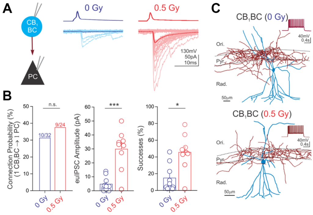
Below is an example of data from paired recordings of hippocampal interneuron (CB­1BC) signaling to pyramidal cells (PC), where CB1BC stimulation evoked inhibitory currents in the PC (panel A). The impact of a manipulation on CB1BC to PC signaling, in this case following proton irradiation (0.5 cGy) was then quantified (panel B). The morphology of the recorded neurons was then reconstructed (panel C). For further information, see Lee S-H et al., 2017, Brain Structure and Function.
Lab Members

Postdoctoral Researcher

Peter Klein

Postdoctoral Researcher

Peter Klein

Peter completed his B.S. in Neuroscience at Bates College and his Ph.D. in Neuroscience with Dr. Mark Beenhakker at the University of Virginia. Since 2008, Peter has focused on investigating how neuronal network activity is perturbed in diseases such as epilepsy. After joining the Soltesz lab in 2018, Peter has been researching how the contributions of specific populations of hippocampal interneurons are altered in epilepsy using mostly in vitro electrophysiology approaches. He is also interested in how neuroimmune interactions contribute to modulating hippocampal excitability following seizures, irradiation, chemotherapy exposures, or other central nervous system insults.

URee Chon has graduated with a B.S. in Biochemistry and Molecular Biology from Pennsylvania State University. As a first year rotating Neuroscience graduate student at Stanford, she is mentored by Dr. Nguyen in the Soltesz lab.

Shreya Malhotra is a first-year medical student at Stanford. In the lab, she will be studying the role of hippocampal circuitry in epilepsy. She will be working closely with Dr. Dudok.

Jesslyn Homidan is a recent graduate of UC Berkeley, where she received her B.A. in Molecular and Cellular Neurobiology. At Berkeley, she worked as an undergraduate apprentice in Dr. Na Ji’s Lab conducting biophysics research on the thalamus and visual cortex. Jesslyn is excited about her new role as a Research Assistant in our lab.

Aaron Milstein has accepted his new position at Rutgers University. He is an Assistant Professor in the Dept. of Neuroscience and Cell Biology and the Dept. of Neurosurgery at Robert Wood Johnson Medical School, and a resident faculty member at the Center for Advanced Biotechnology and Medicine. As an Instructor mentored by Ivan Soltesz, Aaron developed large biologically-detailed neuronal network models to dissect the circuit components of memory and dysfunction in epilepsy.

The Soltesz Team is wishing him all the best in his new career path!

Barna Dudok received a K99/R00 Career Development Award, entitled “Perisomatic inhibition in epilepsy”. He will study the role of a specific type of GABAergic inhibitory interneuron (cholecystokinin-expressing basket cells) in controlling hippocampal network activity in a mouse model of temporal lobe epilepsy using in vivo calcium imaging.

The optical transparency and small brain sizes of larval zebrafish have enabled unprecedented access to single-cell neural dynamics across the whole brain.  With this, we seek to discover novel principles of cellular resolution communication networks that are integral for generating pathological seizure dynamics. Just recently, we have incorporated the chronically epileptic mouse into our analysis. Which is to say, we can now draw insights across evolutionary time, and which will be directly relevant towards translation. Our key advances so far:

  • Built biologically constrained single-cell whole brain effective connectivity models optimized from experimentally acquired calcium dynamics as a platform for hypothesis testing.
  • With the help of novel clustering algorithms, we discovered a new ‘type’ of neuron – the higher-order hub neuron – which is responsible for destabilizing epileptic networks by facilitating unrestrained excitation downstream.
  • Things are surprisingly similar in chronically epileptic mice! This means we have characterized a previously unrecognized organization of epileptic networks that leads us to predict a new target for seizure control.

Moving forward, we will use our lab’s full-scale data-driven models of hippocampal dentate gyrus and CA1 to dig deeper into the underlying circuit mechanisms responsible for the emergence of higher-order hubs in epileptic networks.

Screen Shot 2020-10-02 at 9.18.30 AM

Lab Members

No members found