In: Research

October 13, 2020

Patch-Clamp Recordings

The brain is composed of a wide array of interconnected neurons, including both excitatory principal cells and a diverse assortment of regulatory interneurons. As neurons communicate through electrochemical mechanisms, characterizing the electrical signatures of both control and experimentally perturbed neurons provides great insight into the properties of the fundamental components that compose brain circuits.

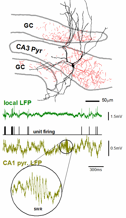
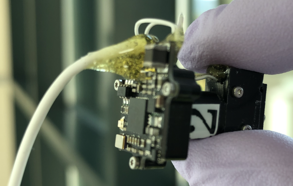
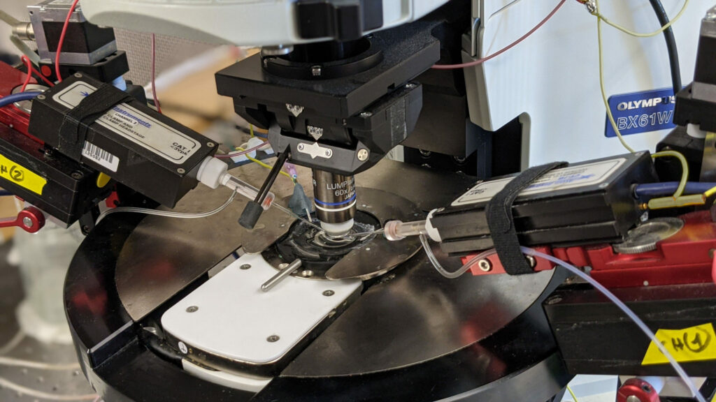
With whole cell electrophysiology, we use finely pointed glass electrodes to precisely record the electrical signal from within individual neurons. Thus, we can examine both the functional electrical properties intrinsic to that particular cell and monitor the synaptic inputs that neuron receives. While recordings are often made from the neuronal cell body, the Soltesz lab has experience recording specifically from smaller structures, including hippocampal dendrites or even axonal terminals.

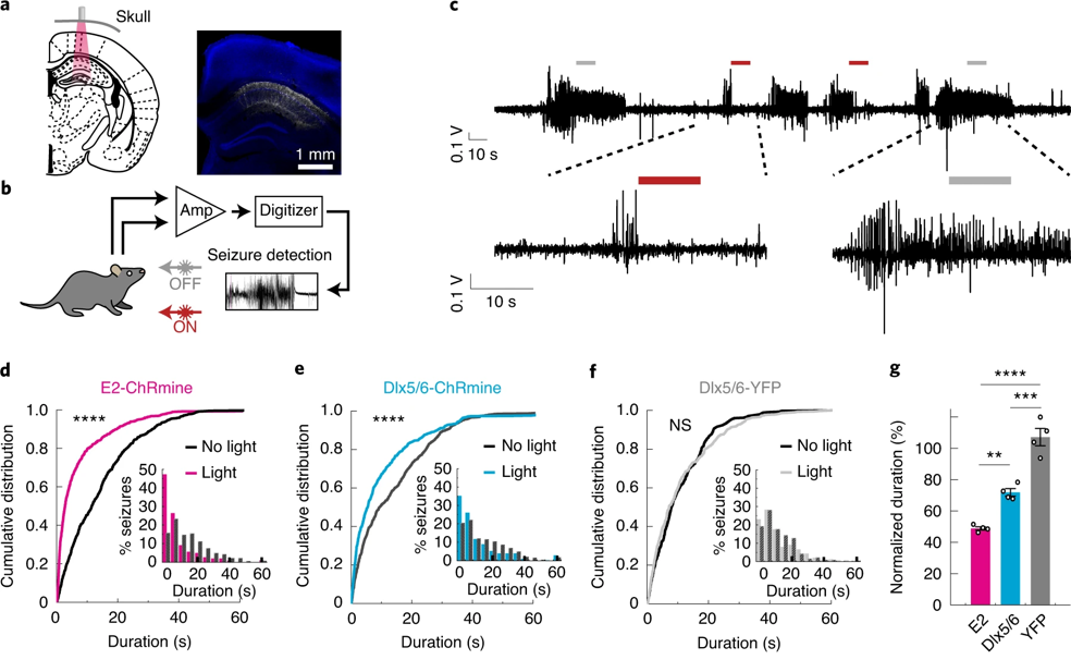
We also utilize several variants on the whole cell electrophysiology approach to interrogate other features of neuronal networks. We can apply compounds to manipulate neuronal activity either through bath application onto the entire brain slice, or through more targeted application via a microinjection system. Through paired patch-clamp recordings, we explore how two simultaneously recorded neurons (whether of the same or different cell types) are interconnected and how they influence the electrical properties of each other. We also employ optogenetic techniques to alter the activity of genetically targeted neuronal populations in response to light stimulation, both to examine their downstream connectivity and how they influence the electrical properties of other neurons. Furthermore, we have the capacity to combine whole cell electrophysiology with simultaneous high-speed imaging of optical probes (e.g. calcium and voltage indicators), enabling us to incorporate the high single cell precision of electrophysiology with broader population level readouts of activity within a brain slice.

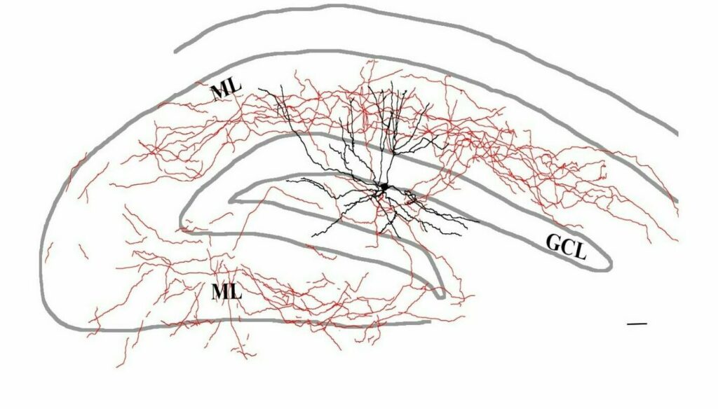
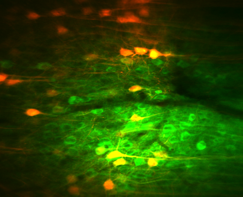
Neurons recorded with whole cell electrophysiology can be filled with intracellular tracers, allowing enabling later morphological reconstruction of the cell. Subsequently, the labelled cell can be further stained for neurochemical markers with immunohistochemistry.

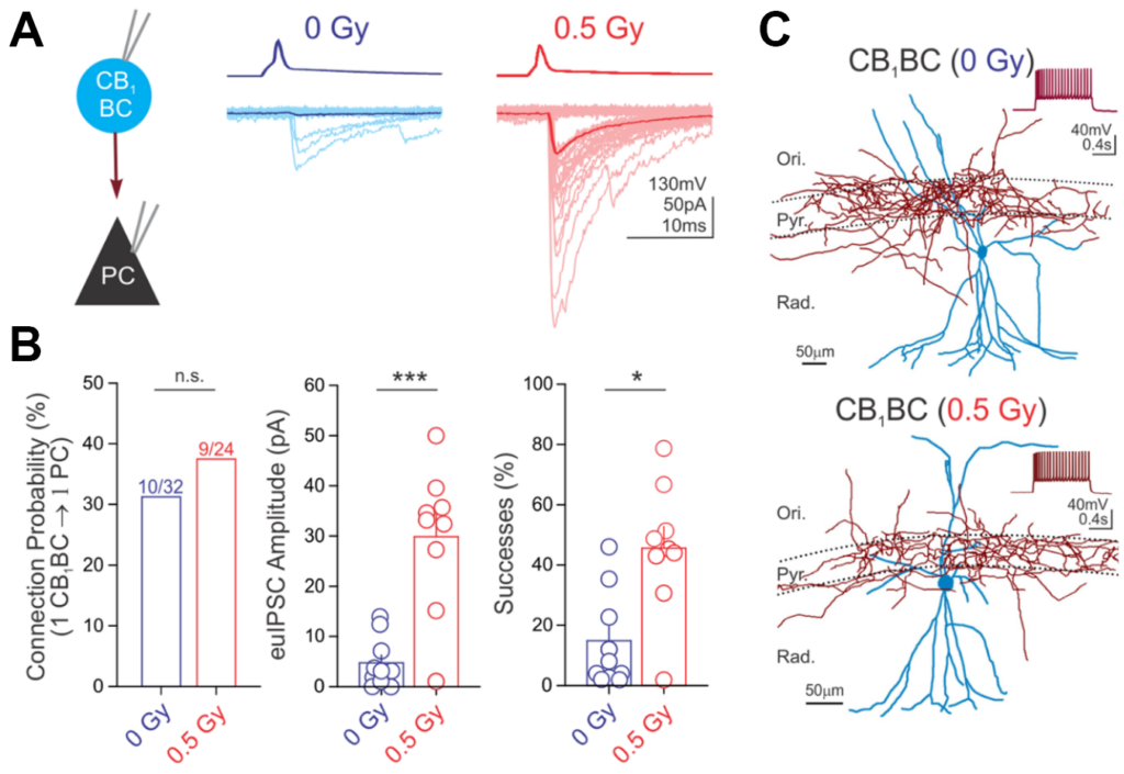
Below is an example of data from paired recordings of hippocampal interneuron (CB­1BC) signaling to pyramidal cells (PC), where CB1BC stimulation evoked inhibitory currents in the PC (panel A). The impact of a manipulation on CB1BC to PC signaling, in this case following proton irradiation (0.5 cGy) was then quantified (panel B). The morphology of the recorded neurons was then reconstructed (panel C). For further information, see Lee S-H et al., 2017, Brain Structure and Function.

Lab Members

Postdoctoral Researcher

Peter Klein

Postdoctoral Researcher

Peter Klein

Peter completed his B.S. in Neuroscience at Bates College and his Ph.D. in Neuroscience with Dr. Mark Beenhakker at the University of Virginia. Since 2008, Peter has focused on investigating how neuronal network activity is perturbed in diseases such as epilepsy. After joining the Soltesz lab in 2018, Peter has been researching how the contributions of specific populations of hippocampal interneurons are altered in epilepsy using mostly in vitro electrophysiology approaches. He is also interested in how neuroimmune interactions contribute to modulating hippocampal excitability following seizures, irradiation, chemotherapy exposures, or other central nervous system insults.

The optical transparency and small brain sizes of larval zebrafish have enabled unprecedented access to single-cell neural dynamics across the whole brain.  With this, we seek to discover novel principles of cellular resolution communication networks that are integral for generating pathological seizure dynamics. Just recently, we have incorporated the chronically epileptic mouse into our analysis. Which is to say, we can now draw insights across evolutionary time, and which will be directly relevant towards translation. Our key advances so far:

  • Built biologically constrained single-cell whole brain effective connectivity models optimized from experimentally acquired calcium dynamics as a platform for hypothesis testing.
  • With the help of novel clustering algorithms, we discovered a new ‘type’ of neuron – the higher-order hub neuron – which is responsible for destabilizing epileptic networks by facilitating unrestrained excitation downstream.
  • Things are surprisingly similar in chronically epileptic mice! This means we have characterized a previously unrecognized organization of epileptic networks that leads us to predict a new target for seizure control.

Moving forward, we will use our lab’s full-scale data-driven models of hippocampal dentate gyrus and CA1 to dig deeper into the underlying circuit mechanisms responsible for the emergence of higher-order hubs in epileptic networks.

Screen Shot 2020-10-02 at 9.18.30 AM

Lab Members

No members found
April 7, 2020

Octopus Research

Cephalopods, including the cuttlefish, octopus, and squid, possess one of the most advanced nervous systems among invertebrates. With their advanced nervous systems, cephalopods are able to perform sophisticated behaviors such as navigating in open water to search for food. Yet how their nervous systems accomplish spatial navigation remains completely unknown. On the other hand, much work has been done in mammals, especially the rodent, to reveal the part of their nervous systems underlying spatial navigation. In particular, neurons in the hippocampus exhibit spatially-selective activity that is thought to provide an internal estimate of animals’ current location. This brings up an interesting question: do cephalopods, with their lineage diverges from the vertebrates more than 500 million years ago, evolve a similar or distinct strategy in their nervous systems to solve the navigation problem? To address this question, this project aims to record neuronal activity in the octopus brain while the octopus perform a spatial navigation task. This work has the potential to reveal essential neuronal mechanism underlying spatial navigation, irrespective of different animal species.

Lab Members

Postdoctoral Researcher

Ernie Hwaun

Postdoctoral Researcher

Ernie Hwaun

Ernie completed his PhD in Neuroscience from the University of Texas at Austin. He is interested in how neurons connect with each other to support cognitive functions such as memory. To tackle this problem, Ernie has been using in vivo extracellular recording techniques to obtain neuronal activity while animals perform a memory task. Besides research, Ernie enjoys playing basketball with friends and reading manga.

December 18, 2019

Juxtacellular Recordings

Pyramidal cells, the principal excitatory cells of the hippocampus, are under the control of a remarkably diverse set of inhibitory interneurons.  These interneurons show substantial differences in their morphology, physiological behavior (spiking activity) and neurochemical signatures. A specific combination of these three characteristic features allows an interneuron to fill a specific niche within the hippocampal network. Therefore, to better understand the function of a given interneuron type, we need to gain information about all these features.

It is possible to achieve all of these goals using a juxtacellular recording & labelling method that we perform on awake, non-anesthetized, head-fixed mice that are running or resting on a spherical treadmill. To record neuronal spiking activity, we first position a small glass electrode adjacent to the cell membrane, thus recording action potentials without perturbing the cell (i.e. juxtaposing the pipette to the cell membrane). Following recording, an intracellular tracer such as neurobiotin can be delivered specifically to that cell, enabling later morphological reconstruction of the neuron. Subsequently, the labelled cell is further stained for neurochemical markers with immunohistochemistry.

The example on the right shows an interneuron recorded and labeled using the juxtacellular technique. Axons (red) are located in the CA3 region of the hippocampus and in the adjacent blades of the dentate gyrus as well. Morphological and immunohistochemical analysis identified this cell as a unique type of axo-axonic cell that simultaneously controls the axon initial segments of CA3c pyramidal cells and dentate granule cells. Spiking activity (black trace) and simultaneous local field potential (green trace) at high temporal resolution reveals that this cell stops firing long before sharp wave-ripples. For further information, see Szabo GG et al., 2017, Cell Reports.

Lab Members

Research Scientist

Gergely Szabo

Research Scientist

Gergely Szabo

Gergely is a Basic Life Research Scientist whose main focus is studying the structure and function of hippocampal inhibitory circuitry and its involvement in learning and memory, utilizing techniques such as electrophysiology, optogenetics, and imaging. Gergely received his MS in Biology from Eotvos Lorand University in Hungary and his Ph.D. in Neuroscience from Semmelweis University in Hungary, after which he joined the Soltesz Lab as a postdoctoral fellow.

December 18, 2019

Behavioral Experiments

A major impediment to progress towards reproducible, rigorous preclinical research in epilepsy is that current experimental approaches often require prohibitively time and labor-intensive 24/7 video-EEG monitoring and inherently subjective scoring of seizures by human observers. In collaboration with the Datta lab at Harvard, who showed that complex animal behaviors are structured in stereotyped modules (“syllables”) at sub-second timescales arranged according to specific rules (“grammar”), we are developing a technique employing video imaging with a 3D camera and artificial intelligence (AI)-assisted video analysis to assess epileptic behavior in an automated and unbiased manner.

Lab Members

Instructor

Tilo Gschwind

Instructor

Tilo Gschwind

Focusing on hippocampal network reorganization in temporal lobe epilepsy while tackling inherent problems of decade-old technology to advance epilepsy research, his project in the Soltesz lab provides an optimal opportunity to contribute to the interdisciplinary discourse between the fields of neuroscience and AI.

We use high-density silicon probes in the hippocampus and related circuits to record from single neurons and monitor the local field potential dynamics with high spatiotemporal resolution. When combined with optogenetics, we can “tag” neurons to achieve cell-type specific recordings as mice navigate a cue-rich treadmill while head-fixed or during restraint-free behavior. Optogenetics also enables us to study how specific cells within and outside of the hippocampus control important LFP dynamics, including theta and sharp-wave ripples. We use a variety of electrode layouts, including Neuropixel probes, to cover a range of research questions.

Lab Members

Postdoctoral Researcher

Ernie Hwaun

Instructor

Tilo Gschwind

Postdoctoral Researcher

Ernie Hwaun

Ernie completed his PhD in Neuroscience from the University of Texas at Austin. He is interested in how neurons connect with each other to support cognitive functions such as memory. To tackle this problem, Ernie has been using in vivo extracellular recording techniques to obtain neuronal activity while animals perform a memory task. Besides research, Ernie enjoys playing basketball with friends and reading manga.

Instructor

Tilo Gschwind

Focusing on hippocampal network reorganization in temporal lobe epilepsy while tackling inherent problems of decade-old technology to advance epilepsy research, his project in the Soltesz lab provides an optimal opportunity to contribute to the interdisciplinary discourse between the fields of neuroscience and AI.

December 18, 2019

Closed-Loop Optogenetics

Optogenetics is a control technology that allows the fast, selective excitation or inhibition of specific neurons with light by expressing light-sensitive proteins (opsins) in particular cell types, enabling causal control of neuronal activity in behaving animals. We have developed a closed-loop, real-time, on-demand system to utilize optogenetics to provide effective seizure control in experimental models of temporal lobe epilepsy (TLE) (Krook-Magnuson et al., 2013) in a spatial, temporal, cell-type and direction of modulation-(excitation or inhibition) selective manner. Importantly, closed-loop optogenetic intervention (COI) was capable not only of curtailing electrographic seizures, but also of significantly decreasing the number of behavioral seizures. COI has proven to be a powerful new tool to understand epileptic circuits, and our lab has used it to directly test the role of the cerebellum as a distant site for seizure control in TLE (Krook-Magnuson et al., 2014) and the role of mossy cells in epilepsy (Bui et al., 2018). One of the most attractive features of COI is that the intervention is highly selective, occurring only when needed (at the time of seizures) and where it is needed, causing selective disruption of seizures while only minimally interfering with ongoing computations necessary for normal brain function. We have recently shown that prolonged application of COI can ameliorate cognitive deficits in TLE (Kim et al., 2019), providing evidence that this technology can improve both seizure burden and the associated comorbidities of epilepsy. Continuing work in the Soltesz lab is focused on utilizing COI as well as incorporating recently developed molecular tools to use with the system to identify and understand epileptic circuits both within and outside of the hippocampus. Ultimately our work aims to provide critical insight into potential targets and avenues for intervention in the treatment of epilepsy.

Lab Members

Instructor

Tilo Gschwind

Instructor

Tilo Gschwind

Focusing on hippocampal network reorganization in temporal lobe epilepsy while tackling inherent problems of decade-old technology to advance epilepsy research, his project in the Soltesz lab provides an optimal opportunity to contribute to the interdisciplinary discourse between the fields of neuroscience and AI.

To monitor the activity of specific cells during unrestricted behavior, we use open-source head-mounted miniscopes developed at UCLA (miniscope.org) to measure calcium activity. The main advantage of this approach over head-fixed 2-photon imaging is the ability to perform a broader range of behavioral tasks including artificial intelligence-based behavioral analysis. Combining these cell type-specific recordings with unbiased sub-second behavioral analysis using AI facilitates the characterization of the cellular underpinnings of behavior at a resolution not possible with conventional approaches.

Calcium Imaging from the Anterior Cingulate Cortex during exploration.

Lab Members

No members found
December 18, 2019

In-vivo Calcium Imaging

We’re using 2-photon microscopy to record neuronal activity with single-cell resolution in the hippocampus of awake, behaving mice. Taking advantage of cell type-specific viral targeting of various biosensors, our goal is to better understand how distinct populations of excitatory and inhibitory neurons are recruited during network oscillations in the healthy brain, and during pathological activity such as seizures in the epileptic brain.

Lab Members

Research Scientist

Gergely Szabo

Research Scientist

Gergely Szabo

Gergely is a Basic Life Research Scientist whose main focus is studying the structure and function of hippocampal inhibitory circuitry and its involvement in learning and memory, utilizing techniques such as electrophysiology, optogenetics, and imaging. Gergely received his MS in Biology from Eotvos Lorand University in Hungary and his Ph.D. in Neuroscience from Semmelweis University in Hungary, after which he joined the Soltesz Lab as a postdoctoral fellow.

December 18, 2019

Computational Modeling

The hippocampal circuits that store and recall spatial information are comprised of diverse cell types, each exhibiting distinct dynamics and complex patterns of synaptic connectivity. Thus, even highly specific experimental perturbations of a single component of these neuronal circuits can have counterintuitive effects on their internal dynamics and output. Computational modeling offers experimentalists a framework to integrate their knowledge, make explicit the assumptions of their conceptual models, and quantitatively predict how each element of a neuronal network is expected to respond to cell type- or projection-specific perturbations. In the Soltesz lab we build computational models in close collaboration with experimentalists, both in the lab, across Stanford, and at other institutions through a multi-site NIH BRAIN Initiative collaboration. Our large-scale network models of the hippocampus are continuously refined to incorporate newly obtained experimental constraints, and numerical simulations are carried out to test hypotheses, compare candidate biophysical and network mechanisms for memory storage and recall, and aide in the interpretation of physiological and behavioral experimental data. The ultimate goal of these efforts is to obtain a deep conceptual understanding of the cellular and network mechanisms that mediate “memory replay” events called sharp-wave ripples by simulating a large-scale model of the hippocampus that reproduces for the observed firing properties of all cell types during sharp-waves.

Lab Members

Research Engineer

Ivan Raikov

Postdoctoral Researcher

Alexandra Chatzikalymiou

Research Engineer

Ivan Raikov

I hold undergraduate and master’s degree in Computer Science from the Georgia Institute of Technology, and a PhD in Biomedical Sciences from the University of Antwerp. I am studying information processing in the hippocampus by means of highly detailed and realistic computational simulation of neuronal networks at 1:1 scale.  More broadly, I am interested in solving the enormous neuroinformatics challenges of computational neuroscience by developing sophisticated computational frameworks capable of expressing, organizing and managing the different types of data and algorithms associated with computational models of neural networks.

Postdoctoral Researcher

Alexandra Chatzikalymiou

Alexandra Chatzikalymniou holds a bachelor’s and a master’s degree in Chemical Engineering from the University of Patras, Greece, and a PhD in Neuroscience and Physiology from the University of Toronto. In her PhD, Alexandra focused on the modelling of theta rhythms using both phenomenological and biophysical models of the rodent hippocampus. As part of her modelling work, she used and analysed state-of-the-art biologically detailed models of the rodent CA1 developed by the Soltesz lab, to understand elements of theta rhythm generation. Alexandra is interested in place cell formation during navigation, and ripple related mechanisms of memory recall and consolidation.